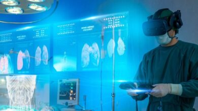
Visualizing the Unseen: Exploring Images of Internal Organs

Have you ever wondered what your internal organs look like? Thanks to medical technology and imaging techniques, we can now visualize the unseen and explore the fascinating world of our own bodies. Images of internal organs provide a unique glimpse into the intricate workings of our systems, allowing doctors to diagnose illnesses and researchers to advance their understanding of human anatomy. In this blog post, we’ll delve into the different types of images available for visualizing internal organs and discuss their functions and uses in modern medicine.
What is an image of an internal organ?
An image of an internal organ is a visual representation of the structure and function of organs within the body. It can provide insights into how different systems are functioning, allowing doctors to diagnose diseases or abnormalities.
There are many types of imaging techniques used to visualize internal organs, including X-rays, CT scans, MRIs, ultrasounds and PET scans.
Internal organs can be incredibly useful in diagnosing conditions like cancer or heart disease. They allow physicians to see any tumors or blockages that might be present in a particular organ system.
Medical imaging has revolutionized medicine by enabling us to “see” inside our bodies without invasive procedures. With these non-invasive tools at their disposal, doctors can make more accurate diagnoses and develop targeted treatment plans for their patients based on precise information about their individual health needs.
Types of images of internal organs
When it comes to visualizing the unseen, medical imaging technology has made great strides in recent years. There are several types of images that can be used to explore internal organs and diagnose ailments.
One common type of image is an X-ray, which uses radiation to capture a two-dimensional image of bones and other dense tissues within the body. This can be useful for identifying fractures or abnormalities in skeletal structures.
Ultrasound is another type of image that uses sound waves to create real-time images of soft tissue structures like organs. It’s non-invasive and relatively inexpensive compared to other imaging techniques.
CT scans use X-rays at different angles around the body to generate detailed 3D images, allowing doctors to see inside organs more clearly than with traditional X-rays alone.
MRI scans use strong magnetic fields and radio waves to produce high-resolution images of internal organs without exposing patients to ionizing radiation from X-rays or CT scans. MRI is particularly useful for examining soft tissues like muscles, tendons, and ligaments.
Each type of image has its own strengths and limitations depending on what needs to be examined. By utilizing these various imaging techniques, doctors are better able to visualize internal organs and provide more accurate diagnoses for their patient’s health concerns.
Functions and uses of images of internal organs
Images of internal organs play a crucial role in the diagnosis and treatment of various medical conditions. One of their primary functions is to help doctors identify, locate, and evaluate abnormalities or diseases present within the body.
These images provide a clear picture of what’s going on inside the patient’s body without requiring invasive procedures such as surgery. They can be used to detect tumors, fractures, blockages, inflammation and other disorders that may not be visible on the surface.
In addition to diagnosis, image Internal organs are also used in treatment planning. For example, if a tumor is detected through imaging tests like MRI or CT scan then radiation therapy can be designed with precision targeting only cancerous cells while minimizing damage to surrounding healthy tissues.
Moreover, these images are also helpful during surgical procedures. Surgeons use them as guides for locating precise areas for incisions and removing abnormal growths or damaged tissue accurately.
Images of internal have revolutionized modern medicine by providing non-invasive methods for diagnosing several health issues. From identifying early-stage cancers to detecting heart disease at its onset – these medical marvels continue to save countless lives every day! Read more…
Conclusion
Images of internal organs have revolutionized the field of medicine. They allow doctors to accurately diagnose and treat diseases while minimizing invasive procedures. There are various types of imaging techniques available today that can provide a clear picture of what is happening inside our bodies.
From X-rays to MRIs, these technologies offer different perspectives and information about organs that would otherwise be impossible to see without surgery.
It’s important for patients to understand how these imaging techniques work and their benefits when being diagnosed or treated for medical conditions. These images often play an essential role in making life-saving decisions.


