Medical imaging exams have revolutionized the way we diagnose and treat diseases. From X-rays to ultrasounds, these technologies provide us with a non-invasive way to get an inside look at our bodies. But did you know that one crucial factor affects the accuracy of medical imaging? That’s right; it’s echotexture! Echotexture refers to how tissue appears on ultrasound images, and it plays a vital role in identifying abnormalities and diagnosing conditions accurately. In this blog post, we’ll explore what homogeneous echotexture means and why it is essential for medical imaging exams. So let’s dive in!
What is Homogeneous Echotexture?
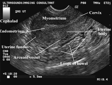
Echotexture is the term used to describe the way tissues appear on ultrasound images. When sound waves penetrate through our body, they reflect back at different rates depending on the type of tissue they encounter. These reflected signals are then captured by an ultrasound machine and translated into an image.
Different tissues have different echogenicity, which means that they reflect sound waves differently. For example, bone appears bright white since it reflects almost all of the sound waves sent its way. In contrast, blood vessels appear as dark lines because they don’t reflect many sound waves.
The homogeneity of echotexture refers to how uniform or consistent a particular tissue’s echo pattern is throughout an image. Homogeneous echotexture occurs when there is little variation in how echoes are produced across a region of interest.
Assessing echotexture is essential for detecting abnormalities such as tumors or cysts since these can disrupt a tissue’s normal echo pattern and create areas with inconsistent texture.
In summary, understanding what echotexture means and how it works is crucial for interpreting medical imaging exams accurately and identifying potential issues early on.
How Does homogeneous Echotexture Affect Medical Imaging?
The echotexture of an organ or tissue is essential in determining its health and diagnosing any abnormalities. In medical imaging exams, such as ultrasounds and MRIs, the echotexture can provide valuable information on the internal structure and composition of the body.
A homogeneous indicates that the tissue or organ has a uniform internal structure with consistent echoes throughout. This is typically associated with good health. On the other hand, an in-echotexture may indicate abnormalities such as tumors, cysts, or inflammation.
For example, liver disease can be detected through changes in echogenicity caused by fatty infiltration or cirrhosis. Similarly, breast cancer often appears as a hypoechoic mass due to its increased density compared to surrounding healthy tissue.
By analyzing the echotexture of organs and tissues during medical imaging exams, doctors can make accurate diagnoses and develop effective treatment plans for their patients.
How to Improve Echotexture in Medical Imaging Exams
Improving echotexture in medical imaging exams is essential for accurate diagnosis and treatment plans. Here are some ways to improve echotexture:
1. Patient Preparation: Before the exam, patients should be adequately hydrated as dehydration can affect tissue density and alter echogenicity.
2. Proper Technique: The transducer should be placed perpendicular to the skin surface with adequate pressure to prevent air from entering between the skin and the transducer.
3. Adjust Settings: Correctly adjusting gain, depth, frequency, dynamic range, and time-gain compensation settings can help improve echotexture.
4. Image Optimization: Optimizing images by adjusting brightness/contrast can enhance the visualization of structures with abnormal or heterogeneous textures.
5. Use Contrast Agents: In cases where it’s challenging to differentiate between tissues due to similar textures (such as liver masses), contrast agents may be helpful in improving image quality.
By following these tips accurately, one can effectively improve their chances of obtaining high-quality homogeneous images that provide clinicians with relevant information needed for better diagnoses and treatment plans. Read more…
Conclusion
Echotexture plays a vital role in medical imaging exams as it helps doctors to diagnose and monitor various medical conditions. Homogeneous echotexture is particularly important because it ensures that the internal structures of organs are uniform and free from abnormalities.
By implementing the tips discussed in this article, such as using high-quality equipment and optimizing patient positioning, healthcare professionals can improve echotexture in their patients’ medical images.
As technology continues to advance in the field of medical imaging, we can expect even greater precision and clarity when it comes to detecting subtle changes within our bodies. By staying up-to-date with new developments and techniques for improving echotexture, we can ensure that patients receive the best possible care for years to come.